On March 6, the team of Professor Li Feng of Orthopedics of Tongji Hospital successfully implemented "lumbar tumor resection + reconstruction surgery" for a lumbar tumor patient with the help of three cutting-edge technologies of 3D printing, mixed reality, and computer-assisted navigation. This is the first case.
Mr. Tian, 39, began to experience recurrent pain in his waist a year ago, and he felt pain after the New Year.
The local hospital MRI showed that there was a tumor on the lumbar spine, the size of the goose egg, and the fifth lumbar spine was eroded, which was near the connection between the lumbar spine and the pelvis.
Through examination, Tongji Hospital's orthopedics confirmed that Tian was a giant cell tumor of the lumbar spine. The fifth lumbar spine was damaged by more than 70%, and the adjacent soft tissue was also damaged.
This kind of tumor is a borderline benign tumor with a malignant tendency. If it is not completely removed, the patient's pain will gradually increase, and even paralysis due to bone collapse. If the tumor cells metastasize, it will be life-threatening.
In the early stage, if the tumor is completely and completely removed as soon as possible and the vertebral body is eroded, the patient is likely to completely defeat the tumor.
After many discussions, the Li Feng team decided to jointly apply "3D printing + mixed reality + computer-aided navigation" to tailor a minimally invasive, personalized, and precise treatment plan for patients.

The first 4 days, according to Mr. Tian's original imaging data, the lesion model printed using 3D printing technology was delivered to Professor Li in time.
Professor Li immediately took the model to the ward to "reproduce" the lesion environment for Mr. Tian and explained the surgical plan in detail.
Mr. Tian said, "I can't understand the film, I can see it at a glance, I can understand it, and I'm not so afraid now."
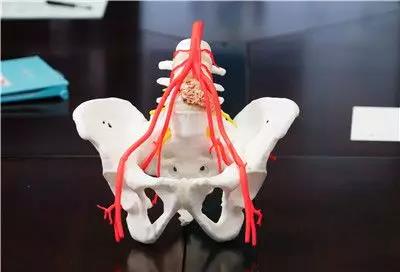
Two days before the operation, a personalized artificial vertebra made by 3D printing was sent to orthopedics. This vertebra was implanted into Mr. Tian's lumbar vertebra after the tumor was removed, replacing the vertebral body eroded by the tumor.
The operation was performed on March 6.
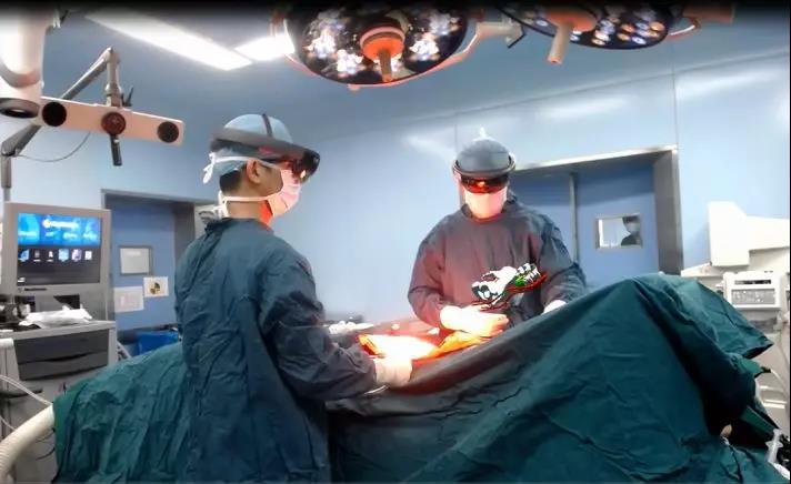
During the operation, Professor Li Feng wore Hei Chao's virtual glasses and used only a few seemingly easy gestures to operate. The virtual 3D images of Mr. Tian's lesion model and artificial vertebral body model were matched with Mr. Tian's real lumbar spine. , More accurate tumor removal.After transforming Mr. Tian's position in general anesthesia, the fifth lumbar spine containing the tumor was finally cut off in one piece.
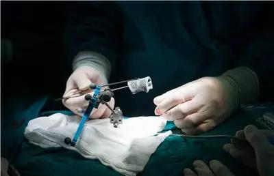
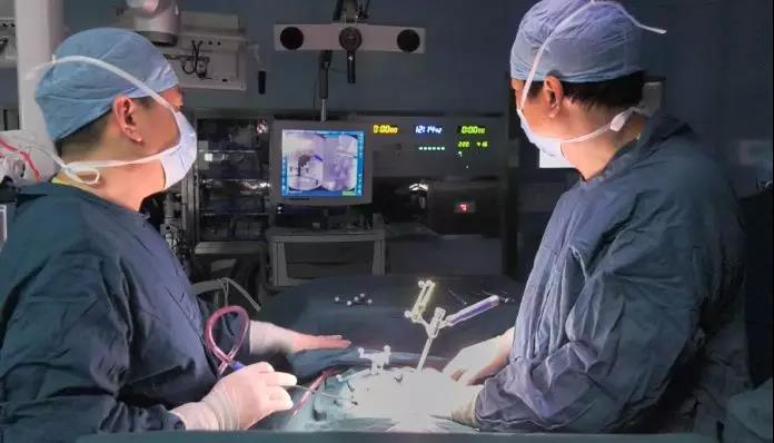
While fixing the artificial vertebra, Li Feng held a needle-like object on Mr. Tian's spine while looking up at the computer screen at the end of the operating table. On the screen was an image of Mr. Tian's spine. To move on the map, and navigate according to the map to find a suitable nail placement.
The operation ended successfully. The tumor and the eroded vertebral body were resected, the artificial vertebral body was firmly fixed at the spinal gap with two orthopedic rods and eight screws, and the spine structure was reconstructed.
Compared with traditional surgical procedures, patients have less trauma, less damage to soft tissues, blood vessels, and nerves, more complete tumor clearance, and lower local tumor recurrence rates.
The patient's vital signs were stable after the operation and returned to the ward. The lesion has been submitted for examination, and further treatment will be guided according to the results of the examination.
At present, the patient is conscious, his back pain is relieved, and he is recovering well.

 Your current location: